Groundbreaking Study on Mouse Brain Challenges Over a Century of Axon Structure Assumptions
2024-12-02
Author: Noah
Introduction
A revolutionary new study from Johns Hopkins Medicine is prompting scientists to rethink long-held beliefs about the shape of axons, the threadlike structures that facilitate communication between brain cells. For over a century, biology texts have illustrated axons as uniform cylindrical tubes, but this groundbreaking research suggests a far more complex reality.
Key Findings
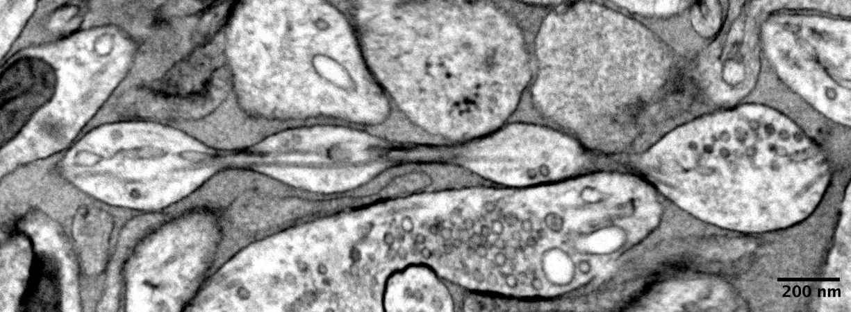
The findings, published on December 2 in Nature Neuroscience, reveal that axons may actually resemble a string of pearls rather than the simplistic tube-like structures that have dominated educational materials. Shigeki Watanabe, Ph.D., an associate professor of cell biology and neuroscience at Johns Hopkins, emphasized the importance of this understanding, noting that 'Axons are the cables that connect our brain tissue, enabling learning, memory, and other functions.'
Challenging Previous Assumptions
Interestingly, previous knowledge established that such pearl-like formations, referred to as axon beading, commonly appear in dying brain cells and are linked to neurodegenerative diseases like Parkinson's. However, this new study suggests these structures exist under healthy conditions as well, challenging previous assumptions about neuron morphology.
Research Methodology
Watanabe's curiosity about axon beading was sparked during a discussion with Swiss scientist Graham Knott, leading to a series of experiments to dissect the intricate physical properties of axons. The research team utilized cutting-edge high-pressure freezing electron microscopy, allowing them to capture the nanoscale structures of axons without losing their shape — akin to freezing a grape rather than dehydrating it into a raisin.
Diverse Neuron Analysis
The study analyzed various types of mouse neurons: those cultivated in a lab, adult mouse neurons, and embryonic neurons, all of which were found to exhibit the pearl shape. The researchers introduced the term 'non-synaptic varicosities' to describe these swellings along the axons.
Mathematical Insights
Mathematical modeling played a crucial role in the study, revealing that the mechanical properties of axonal membranes significantly influence the formation of these structures. By adjusting sugar concentrations and the tension within the axonal membranes, the size of the pearl-like formations could be altered. Removing cholesterol from the membrane was also shown to reduce pearling but adversely affected the axon's electrical signaling capabilities.
Implications for Neural Function
Furthermore, applying high-frequency electrical stimulation caused the swollen structures to expand, improving the speed of electrical impulse transmission through the axons. However, when cholesterol was depleted, the pearls reverted to their original state, highlighting the importance of membrane composition in neural function.
Future Research Directions
Looking ahead, this pioneering research team aims to explore similar axonal structures in human brain tissue, with consent from individuals undergoing brain surgery or who have succumbed to neurodegenerative conditions. This next phase could reveal even greater insights into human brain health and disease, potentially reshaping our understanding of neuronal communication and signaling.
Conclusion
Stay tuned as researchers continue to unravel the complexities of the brain, one pearl at a time!


 Brasil (PT)
 Canada (EN)
 Chile (ES)
 España (ES)
 France (FR)
 Hong Kong (EN)
 Italia (IT)
 日本 (JA)
 Magyarország (HU)
 Norge (NO)
 Polska (PL)
 Schweiz (DE)
 Singapore (EN)
 Sverige (SV)
 Suomi (FI)
 Türkiye (TR)