Revolutionary Microscopy Breakthrough: Hair-Thin Optical Fiber Unlocks New Frontiers in Medical Imaging!
2024-12-03
Author: Michael
In a groundbreaking advancement, researchers at the University of Adelaide have teamed up with an international team to develop a pioneering method that enables advanced microscopy through an optical fiber thinner than a human hair. This innovative technology holds the promise of transforming medical imaging and diagnostics.
Dr. Ralf Mouthaan, a key player at the University of Adelaide's Center of Light for Life, emphasized the significance of the breakthrough. "Recent advances in optics have allowed the controllable delivery of light through ultra-thin fibers; however, creating complex light patterns essential for advanced microscopy has remained a challenge until now," he stated.
With the new approach, microscopic images can be gathered from areas of the human body that were previously difficult, if not impossible, to examine. This development significantly minimizes the risk of tissue damage, offering a less invasive alternative to traditional imaging techniques.
As light traverses through an optical fiber, it often becomes distorted. Particularly when the fiber's diameter nears that of a human hair, this distortion can result in chaotic, granular patterns. However, recent innovations have managed to correct these distortions, allowing ultra-thin devices to venture into uncharted territories of the human anatomy.
This breakthrough paves the way for advanced microscopy techniques such as light sheet microscopy, a method that creates volumetric images by capturing one plane at a time, and stimulated emission-depletion (STED) microscopy, capable of imaging structures at the nanoscale—just a billionth of a meter in diameter.
The collaborative project also includes contributions from experts at the University of Nottingham and the University of Cambridge. Collectively, they have demonstrated the ability to pre-shape light to generate highly specific optical patterns, overcoming the issues posed by distortion.
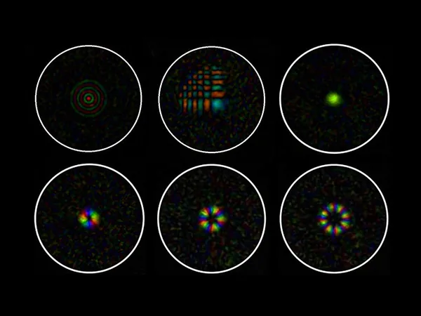
Their findings, recently published in *Advanced Optical Materials*, reveal remarkable control over the amplitude, phase, and polarization of light at the fiber’s output. The researchers showcased the projection of specialized light patterns, including Bessel beams, Airy beams, and Laguerre-Gaussian beams—each serving unique roles in modern microscopy.
Dr. Mouthaan highlighted the revolutionary implications of this technology, stating, "While many cutting-edge microscopes require vast laboratory space, this approach is a monumental advancement toward miniaturizing microscopes to the point where imaging can occur inside the human body. The applications are virtually limitless!"
Excitingly, the Adelaide team is gearing up to demonstrate the first proof of concept for "endomicroscopes," while their collaborators in Nottingham are focused on creating an endoscope tailored for clinical applications.
This innovative technique not only enhances the capabilities of microscopy but could revolutionize the way medical professionals diagnose and treat patients, potentially leading to earlier and more accurate diagnoses. The future of medical imaging is indeed bright—what other secrets could lie just beneath the surface, waiting to be uncovered?


 Brasil (PT)
 Canada (EN)
 Chile (ES)
 España (ES)
 France (FR)
 Hong Kong (EN)
 Italia (IT)
 日本 (JA)
 Magyarország (HU)
 Norge (NO)
 Polska (PL)
 Schweiz (DE)
 Singapore (EN)
 Sverige (SV)
 Suomi (FI)
 Türkiye (TR)