Incredible! Baby Born with Giant Red Balloon on His Back Defies Odds After Rare Surgery!
2025-01-09
Author: Kai
Introduction
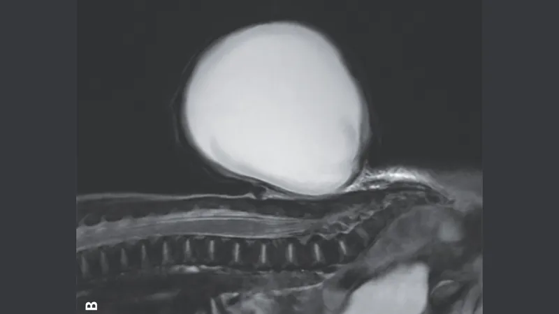
In a remarkable case that has caught the attention of the medical community, a newborn infant developed a striking giant red sac, akin to a balloon, protruding from his lower back due to a condition known as spina bifida. Captured by doctors at Massachusetts General Hospital in Boston, this unusual occurrence highlights both the resilience of medical science and the importance of prenatal care.
Understanding Spina Bifida
The protruding sac measured approximately 3 inches (7.7 cm) in length, 2.8 inches (7.1 cm) in width, and 2.1 inches (5.3 cm) in depth. This phenomenon is a result of a neural tube defect, which ranks as the second most prevalent congenital disability, impacting between 5 to 8 infants in every 10,000 births in the United States.
To better understand the defect, it's essential to know that the neural tube is a crucial structure that forms during the early stages of pregnancy—the third and fourth weeks post-conception—which eventually develops into the brain and spinal cord. In some instances, this process does not proceed correctly, resulting in an opening in the spine. Often, this gap is concealed by skin, causing no symptoms and going unnoticed. However, in rare cases, such as this one, fluid and tissue enveloping the spinal cord can protrude through the gap, forming a sac, leading to a specific type of spina bifida known as meningocele.
Causes and Risk Factors
While the exact causes of spina bifida remain elusive, experts suggest that a combination of genetic, nutritional, and environmental factors can heighten the risk. For instance, inadequate intake of folate or vitamin B9 during the early stages of pregnancy, certain medications, and poorly managed diabetes are notable risk factors. Interestingly, in this case, the baby's condition did not stem from any of those common risks, as confirmed in a report published in the prestigious *New England Journal of Medicine*.
Detection and Treatment
The spinal defect was first detected during a routine ultrasound at approximately 20 weeks of gestation. Children diagnosed with meningoceles typically encounter mild issues such as bladder or bowel problems, but the condition is usually manageable with surgical intervention, which can occur either in utero or after birth.
In more severe cases, such as myelomeningocele, where neural tissue is also involved in the sac, serious complications like paralysis, recurrent urinary tract infections, and meningitis may arise. Fortunately, the baby in this story underwent successful surgery to remove the sac and reconstruct the spinal cord shortly after birth. Remarkably, just four days post-surgery, he was discharged home, and during his six-month follow-up, physicians reported that he was developing well without complications.
Conclusion
This inspiring story serves as a reminder of the advances in prenatal care and pediatric medicine, illustrating how swift medical intervention can make a significant difference in a newborn's life. As awareness of spina bifida grows, expectant mothers are encouraged to prioritize prenatal vitamins and healthcare visits, which can dramatically enhance outcomes for their babies.
 Brasil (PT)
 Canada (EN)
 Chile (ES)
 Česko (CS)
 대한민국 (KO)
 España (ES)
 France (FR)
 Hong Kong (EN)
 Italia (IT)
 日本 (JA)
 Magyarország (HU)
 Norge (NO)
 Polska (PL)
 Schweiz (DE)
 Singapore (EN)
 Sverige (SV)
 Suomi (FI)
 Türkiye (TR)
 الإمارات العربية المتحدة (AR)