Breakthrough in Cancer Diagnosis: Portable Multiphoton Microscopy Revolutionizes Pathology
2025-04-07
Author: Sarah
In an exciting advancement for cancer diagnostics, researchers have developed a portable multiphoton microscopy system that promises to deliver pathology-quality images in real-time, revolutionizing the way biopsies are analyzed. Utilizing cutting-edge femtosecond laser pulses, this innovative imaging technique allows for label-free and damage-free visualization of biological tissues, potentially transforming cancer diagnosis and personalized medicine.
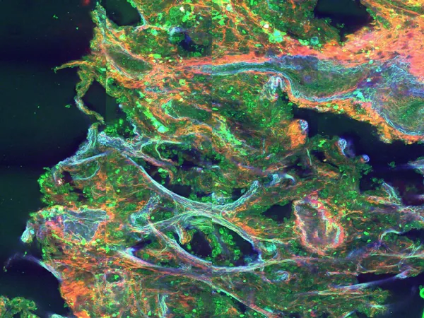
Traditional imaging methods, such as confocal microscopy, often require labor-intensive sample preparation, including slicing and staining, to create tissue contrast. In contrast, multiphoton microscopy leverages nonlinear optical processes to reveal critical molecular and structural details within tissues while keeping samples intact. This unique approach can significantly streamline the diagnostic process, much to the advantage of clinicians and patients alike.
Flash Pathology's Innovative Solution
A standout player in this innovative field is the Netherlands-based startup Flash Pathology, which is diligently working to bring a compact version of multiphoton microscopy to clinical settings. The brainchild of Marloes Groot from Vrije Universiteit Amsterdam, Flash Pathology aims to make high-quality diagnostic imaging accessible in various medical environments. Groot emphasized the power of this technique, expressing her vision of transforming rigorous lab setups into a mobile device that practitioners can use right at the point of care.
Alongside co-founder and Chief Technology Officer Frank van Mourik, Groot successfully compacted the multiphoton device into a transportable system measuring just 60 x 80 x 115 cm. Designed for practical use, the device can be easily wheeled through hospital corridors and delivers images within moments of being set up. Remarkably, recent advancements allow for imaging at dramatically reduced laser power, dropping from 200 mW to just 5 mW without affecting tissue integrity.
Impact on Lung Cancer Diagnostics
The implications of this technology are particularly significant in the realm of lung cancer diagnostics, where rapid analysis of biopsy samples can dictate patient management decisively. Current histopathological techniques often require days to yield critical results, whereas Flash Pathology’s multiphoton microscope can generate detailed images of biopsies as quickly as six minutes post-excision, achieving an impressive 87% diagnostic accuracy rate. This rapid feedback can be crucial for determining whether further biopsies are necessary, significantly cut down on the need for repeated procedures, and ultimately improve patient outcomes.
Advanced Imaging Capabilities
The system operates by generating multiple nonlinear signals concurrently with a single ultrafast femtosecond laser, enabling a more comprehensive diagnostic picture. For instance, second-harmonic generation can detect non-centrosymmetric structures like collagen, while third-harmonic generation elucidates boundaries in cells. This multi-faceted imaging power allows clinicians to observe vital cellular structures and details that pathologists traditionally examine, enhancing diagnostic precision.
Additionally, the performance of multiphoton microscopy derives from the unique capabilities of femtosecond lasers. These ultrashort pulses enhance the likelihood of photon overlap occurring at focal points, significantly boosting the potential for two- and three-photon processes. VALO Innovations' cutting-edge femtosecond lasers can deliver pulses as short as 30 femtoseconds, adapting to the demands of advanced microscopy with high peak power and minimal sample heating.
Future Aspirations and Developments
As Flash Pathology continues to refine its technology, the team is conducting tests in various hospitals, including Amsterdam UMC and the Princess Maxima Center. Their goal is not just to produce a functioning prototype but to develop a certified medical device that integrates artificial intelligence, enabling automated image interpretation and diagnostics.
Looking forward, the potential for this multiphoton microscopy system to revolutionize tissue analysis during surgical procedures is immense. With the ability to provide immediate, in situ diagnoses, this innovative imaging technique stands to greatly enhance patient care and streamline clinical workflows. The promise of having such advanced diagnostic capabilities at the bedside represents a significant leap forward in the fight against cancer.


 Brasil (PT)
 Canada (EN)
 Chile (ES)
 Česko (CS)
 대한민국 (KO)
 España (ES)
 France (FR)
 Hong Kong (EN)
 Italia (IT)
 日本 (JA)
 Magyarország (HU)
 Norge (NO)
 Polska (PL)
 Schweiz (DE)
 Singapore (EN)
 Sverige (SV)
 Suomi (FI)
 Türkiye (TR)
 الإمارات العربية المتحدة (AR)