Breakthrough Study Links Pericoronary Adipose Tissue Features to Coronary Artery Disease Risk: What You Need to Know!
2024-12-19
Author: John Tan
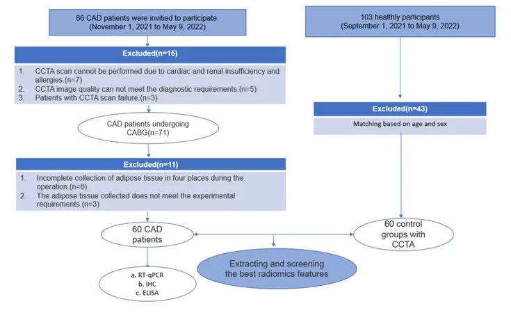
A recent groundbreaking study has unveiled a crucial connection between pericoronary adipose tissue (PCAT) radiomics features and the risk assessment of coronary artery disease (CAD), aided by insights from gene expression analysis. This research, undertaken between November 2021 and May 2022, involved patients who underwent coronary artery bypass grafting (CABG) and sheds light on potential biomarkers for CAD.
Who Was Involved?
The study involved 60 patients diagnosed with CAD, who were carefully selected based on stringent inclusion criteria, including age over 18, complete clinical data, and the absence of cardiac or renal insufficiencies and malignancies. The PCAT, a fat depot surrounding the coronary arteries, was sampled to facilitate an innovative analysis linking its radiomics features to the disease's underlying mechanisms.
CCTA Scanning Technique
All patients underwent advanced coronary computed tomography angiography (CCTA) scans using state-of-the-art 320-row CT devices. This non-invasive imaging technique is pivotal since it allows for a detailed examination of coronary arteries and surrounding fat tissues, providing essential data that can help predict disease risk.
Evaluating Image Quality
The quality of CCTA images was rigorously assessed using both objective and subjective methods, ensuring that critical visual data was reliable for subsequent analysis. The precision of imaging is vital as it directly influences feature extraction and the reliability of the findings.
Radiomics Features Extraction
A total of 1688 radiomics features were extracted from the initial images, with 189 carefully selected based on their strong correlation with CAD. This focused approach highlights the most predictive indicators, enhancing the understanding of CAD at the molecular level. Moreover, an innovative AI tool, ‘CoronaryDoc’, was utilized to automate feature extraction, showcasing the integration of technology in medical research.
Gene Expression Analysis
To further corroborate findings, gene expression levels were assessed in the adipose tissue samples collected. Techniques such as RT-qPCR and immunohistochemistry clearly indicated heightened levels of inflammatory markers like MCP-1 and leptin in PCAT compared to other fat depots like epicardial adipose tissue (EAT) and subcutaneous adipose tissue (SAT). These markers are known to play crucial roles in inflammation and atherosclerosis, further bridging the gap between imaging data and biological pathways.
Significance of Findings
Remarkably, the study concluded that the expression of inflammatory factors and vascular remodeling markers in PCAT could serve as a non-invasive detection method for CAD. This approach not only unveils potential biomarkers but also provides new insights into atherosclerosis mechanisms, demonstrating that molecular features can be deduced from imaging data.
What’s Next?
Although the current study utilized a small sample size, it opens doors for multidisciplinary research aimed at validating these findings across larger populations. Future studies could transform how we assess CAD risk non-invasively, integrating radiomics features with clinical biomarkers for more personalized patient management.
Conclusion
This pioneering research signifies a monumental step towards understanding CAD risk through imaging biomarkers derived from PCAT. The implications for clinical practices are profound, potentially leading to earlier diagnoses and targeted therapy strategies. As we eagerly await further validations and studies, this compelling evidence encourages healthcare professionals to consider the role of radiomics features in enhancing CAD risk assessment and management decisively.
Stay tuned for updates as this fascinating field of research continues to evolve!


 Brasil (PT)
 Canada (EN)
 Chile (ES)
 Česko (CS)
 대한민국 (KO)
 España (ES)
 France (FR)
 Hong Kong (EN)
 Italia (IT)
 日本 (JA)
 Magyarország (HU)
 Norge (NO)
 Polska (PL)
 Schweiz (DE)
 Singapore (EN)
 Sverige (SV)
 Suomi (FI)
 Türkiye (TR)
 الإمارات العربية المتحدة (AR)