Groundbreaking Discovery: Netrin1's Surprising Role in Spinal Cord Formation Revealed!
2024-11-18
Author: Jia
In an astonishing breakthrough, researchers have identified a previously unknown function of Netrin1, a protein already well-known for its role in guiding axon development during embryonic growth. Recent findings from scientists at the Eli and Edythe Broad Center for Regenerative Medicine and Stem Cell Research at UCLA reveal that Netrin1 also plays a crucial role in organizing the developing spinal cord, challenging long-standing assumptions in developmental biology.
Published in the prestigious journal Cell Reports under the title “Netrin1 patterns the dorsal spinal cord through modulation of Bmp signaling,” the study provides new insights that could transform our understanding of how complex spinal circuits form during embryonic development. According to lead researcher Dr. Samantha Butler, a neurobiology professor at UCLA, this discovery stemmed from a curious observation that made them question their previous knowledge about Netrin1.
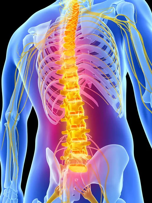
The dorsal spinal cord is essential for processing sensory information such as touch and pain, and its proper development hinges on precise spatial organization. This is where BMP (Bone Morphogenetic Protein) signaling comes into play—an essential communication pathway that must remain confined to specific regions of the spinal cord to ensure proper sensory neuron formation. The study revealed that Netrin1 acts as a critical boundary keeper for BMP signaling, preventing it from spreading to adjacent regions and thus ensuring the organized development of neural networks.
"Without the regulatory influence of Netrin1, the delicate architecture of the neural network could fall into chaos, impacting sensory axon targeting," noted Sandy Alvarez, a graduate student and the study's first author. This underscores the importance of molecular signaling specificity in neural development.
What astonished the team was how Netrin1 functions more like an adhesive surface for axon growth, directly guiding neuronal pathways rather than merely serving as a distant signaling cue. Through innovative experiments in chicken and mouse embryos, as well as mouse embryonic stem cells, researchers traced the movements of Netrin1 within the spinal cord, uncovering a troubling trend: certain nerve cells disappeared when Netrin1 levels increased.
"The initial results were perplexing," Alvarez commented. "We were witnessing the suppression of BMP activity by Netrin1, an interaction that had been largely overlooked in existing scientific literature."
By manipulating Netrin1 levels, the research team demonstrated a corresponding change in the populations of nerve cells in the dorsal spinal cord. Increasing Netrin1 levels led to the loss of specific nerve cell populations, while their removal resulted in an expansion of those populations.
Through rigorous bioinformatics analyses, the team further established that Netrin1 exerts its influence by modulating RNA translation, effectively dampening BMP activity.
Dr. Butler emphasized the vast potential of Netrin1 in neuronal repair, stating, "Given its remarkable capacity to shape neural circuits, our future research will investigate how we can utilize Netrin1 to aid in repairing nerve damage or spinal cord injuries in patients."
The implications of this study stretch far beyond spinal cord development; Netrin1 and BMP also play vital roles in numerous organs throughout the body, making it imperative to reassess their interactions in various physiological contexts. "Our findings prompt a fresh evaluation of how Netrin1 and BMP may be involved in cancer types and developmental disorders," Alvarez concluded.
This remarkable study opens up exciting new avenues for scientific exploration and holds out the promise of significant advancements in regenerative medicine and our understanding of neural disorders. Stay tuned as we watch this groundbreaking research unfold!


 Brasil (PT)
 Canada (EN)
 Chile (ES)
 España (ES)
 France (FR)
 Hong Kong (EN)
 Italia (IT)
 日本 (JA)
 Magyarország (HU)
 Norge (NO)
 Polska (PL)
 Schweiz (DE)
 Singapore (EN)
 Sverige (SV)
 Suomi (FI)
 Türkiye (TR)