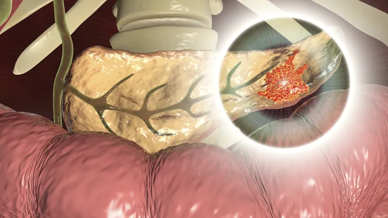
Groundbreaking MRI Technique Identifies Pre-Malignant Pancreatic Cancer Lesions!
2024-12-13
Author: Sarah
Introduction
In an exciting breakthrough, researchers from the Champalimaud Foundation in Lisbon, Portugal, have made significant strides in the early detection of pancreatic cancer lesions. For the first time ever, the study published in Investigative Radiology reports that diffusion tensor imaging (DFI), an advanced MRI technique, has been successfully used to identify pre-cancerous lesions in the pancreas known as pancreatic intraepithelial neoplasia (PanIN).
The Challenge of Detection
Pancreatic cancer, notorious for its late diagnosis, often presents symptoms such as stomach pain, weight loss, and new-onset diabetes, which can easily be mistaken for other health problems. Traditional imaging techniques fail to detect the early stages of this dangerous disease, leaving a critical gap in the ability to diagnose and understand its progression. This new capability using DFI could revolutionize the approach to pancreatic cancer by enhancing the detection of these vital pre-malignant lesions and shedding light on the biological pathways leading to pancreatic ductal adenocarcinomas (PDAC).
Research Insights
"Current imaging modalities don't diagnose PanINs. There is an urgent necessity for better imaging methods to understand and characterize these lesions," said the research team. Given that most cases of PDAC originate from PanINs, which can develop silently for years, the inability to spot these precursor lesions has hindered both research efforts and the development of early-stage treatments.
The DTI Technique
To tackle this challenge, the researchers employed diffusion tensor imaging, a technique that has been used for studying brain tissue for over three decades. Dr. Noam Shamesh, the lead investigator, and head of the preclinical MRI lab at Champalimaud Research, shared, "Although it's not a new method, it was simply never applied to pancreatic cancer precursor lesions. It was Dr. Carlos Bilreiro who approached me with this innovative idea."
How DTI Works
The DTI method evaluates the diffusion of water molecules in tissues. Variations in these movements reveal critical details about the tissue's microstructure, which changes in the presence of PanINs. By utilizing DTI on pancreatic tissues from genetically engineered mice predisposed to develop PanINs, researchers captured high-resolution images that outperformed traditional histological samples in detecting these pre-cancerous conditions.
Clinical Trials and Future Directions
Building on these encouraging findings, the team conducted tests on human pancreatic tissue samples and found that DTI was equally effective in identifying lesions in humans. "Our promising results could significantly enhance diagnostic capabilities," Dr. Shamesh stated.
Challenges Ahead
While the implications of this research are thrilling, the pathway to clinical application remains challenging. The primary obstacle is that standard MRI scanners in clinics typically produce lower-resolution images. Nevertheless, the researchers are determined to address this limitation, with plans for clinical trials to validate DTI in human patients. They are optimistic about the potential to refine DTI for clinical use and could even consider integrating it with innovative approaches like liquid biopsies or artificial intelligence for increased accuracy and specificity in detecting PanINs.
Conclusion
In summary, this pioneering research marks a critical turning point in the fight against pancreatic cancer. As scientists continue to push the boundaries of medical imaging, we may soon see a future where early detection transforms outcomes for patients facing this aggressive disease. Stay tuned for updates on this revolutionary development!


 Brasil (PT)
 Canada (EN)
 Chile (ES)
 España (ES)
 France (FR)
 Hong Kong (EN)
 Italia (IT)
 日本 (JA)
 Magyarország (HU)
 Norge (NO)
 Polska (PL)
 Schweiz (DE)
 Singapore (EN)
 Sverige (SV)
 Suomi (FI)
 Türkiye (TR)