Groundbreaking Ultrasound Technique Reveals Cells and Capillaries in Live Organs – A Game Changer for Medical Imaging!
2025-04-03
Author: Ming
In an exciting development, a collaborative team of researchers from the University of Technology Delft, the Netherlands Institute for Neuroscience, and Caltech has unveiled a revolutionary microscopy technique that harnesses the power of ultrasound to visualize capillaries and cells within living organs. This groundbreaking study has been published in the prestigious journal Science, marking a significant milestone in medical imaging.
Until now, ultrasound has been a staple in clinical settings—most commonly seen in pregnancy scans—but its application for imaging microscopic structures such as cells has remained largely unexplored. "While traditional clinical ultrasound captures real-time images of larger body parts, the inner workings of our bodies at the cellular level have eluded us," stated first author Baptiste Heiles.
Innovative 3D Cell Imaging
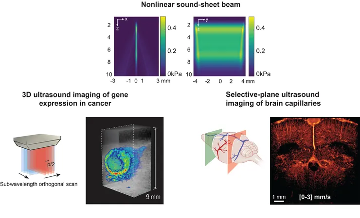
For the first time, the research team successfully employed ultrasound to image specifically labeled cells in three dimensions, within entire organs that are roughly the size of a sugar cube. This advancement is particularly noteworthy since current light-based microscopy methods necessitate the use of non-living samples. As Heiles explained, this previously meant removing the organ from the body, resulting in the loss of valuable data on cell behavior over time.
Light sheet microscopy, the frontrunner in 3D imaging of living cells, is confined to thin or translucent specimens. In contrast, ultrasound can penetrate several centimeters into opaque mammalian tissues, enabling non-invasive imaging of whole organs. Senior researcher David Maresca emphasized that this capability allows scientists to observe living cells in their natural environment—an analysis that light-based strategies cannot perform on larger, intact tissues.
Revolutionizing Imaging with Sound-Reflecting Probes
At the heart of this ultrasonic innovation, termed nonlinear sound sheet microscopy, lies the groundbreaking discovery of a sound-reflecting probe crafted in the Shapiro Lab at Caltech. According to Heiles, this nanoscale gas-filled vesicle enhances ultrasound images, thereby illuminating cells for observation. Engineered with a protein shell, these vesicles can be adjusted for optimal brightness in imaging. Curious about its applications, the researchers have already utilized these gas vesicles to track cancer cells, opening up new horizons in oncology.
Advancements in Brain Imaging
Moreover, the research team has successfully employed ultrasound alongside microbubbles as probes moving through the bloodstream to identify brain capillaries. "To our knowledge, nonlinear sound sheet microscopy stands as the first tool capable of observing capillaries in living brains," Heiles noted. This groundbreaking technique has the potential to diagnose small vessel diseases in patients effectively. Given that microbubble probes are already approved for use in humans, this innovation could find its way into hospitals within just a few years.
A Major Breakthrough for Cancer Research
Beyond immediate clinical applications, this ultrasound technology holds promise for significantly advancing biological research, particularly in the realm of cancer treatment. Maresca remarked, "Our imaging technique not only differentiates healthy tissue from cancerous tissue but can also visualize the necrotic core of tumors—hypoxic regions where cell death occurs. This capability could be vital for monitoring cancer progression and evaluating treatment responses."
This groundbreaking approach to imaging live tissues not only enhances our understanding of cellular dynamics in health and disease but also paves the way for tailored therapeutic interventions. The implications are vast, potentially transforming how we diagnose and treat a variety of conditions, including cancer and vascular diseases. The promise of this innovative ultrasound technology heralds a new era in medical imaging, with the potential to profoundly impact patient outcomes in the near future.

 Brasil (PT)
 Canada (EN)
 Chile (ES)
 Česko (CS)
 대한민국 (KO)
 España (ES)
 France (FR)
 Hong Kong (EN)
 Italia (IT)
 日本 (JA)
 Magyarország (HU)
 Norge (NO)
 Polska (PL)
 Schweiz (DE)
 Singapore (EN)
 Sverige (SV)
 Suomi (FI)
 Türkiye (TR)
 الإمارات العربية المتحدة (AR)