Revolutionary Cardiac MRI Strategy Promises Breakthrough in Complex Tachycardia Treatment!
2024-10-08
Author: Wei
Introduction
In an exciting advancement for cardiac care, researchers have unveiled a groundbreaking strategy that could transform the treatment of complex tachycardias through enhanced cardiac MRI (Magnetic Resonance Imaging) techniques. A multicenter study, spearheaded by experts from Hospital Clínico San Carlos and the Centro Nacional de Investigaciones Cardiovasculares in Madrid, has successfully validated this innovative approach, which could hold the key to improving the efficacy of ablation procedures for patients suffering from these potentially life-threatening arrhythmias.
Ablation Procedures and Their Importance
Ablation procedures, crucial in the management of ventricular tachycardias—often triggered by scarring from previous heart attacks—typically involve the application of either heat or cold to targeted heart tissue. The coordinated efforts of leading institutions both in Spain and the Netherlands, including Hospital Universitario de la Paz and Maastricht University Medical Center, are paving the way for this clinical evolution.
Innovative Image Processing Methods
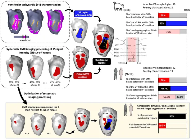
This groundbreaking study, published in the esteemed journal "Europace," introduces advanced image processing methods that aim to accurately identify problematic heart regions—particularly those affected by post-infarction scarring that can sustain ventricular tachycardia. By streamlining the imaging process, researchers have found a way to minimize biases, enhancing the detection of areas of concern for arrhythmia, and ultimately preparing for more effective preoperative planning.
Research Findings and Implications
Utilizing a swine model of myocardial infarction, the research team analyzed how variations in imaging parameters often led to misidentifications of crucial cardiac tissue. The resulting strategy—validated through a comprehensive multicenter study conducted from 2013 to 2022—has shown to improve the precision of identifying these critical circuits in human patients.
Expert Insights
David Filgueiras, the study coordinator and cardiologist, stated, "Our findings address important gaps in imaging data integration for ablation procedure planning. This innovative approach holds the potential to drastically reduce procedure duration and complications—especially in patients who suffer from severe, poorly tolerated tachycardia episodes. Remarkably, we can achieve high success rates without invasive catheter mapping, which is often deemed too risky."
Advancements in Imaging Capabilities
By deepening visualization capabilities to examine cardiac tissue at various depths, this new strategy allows for a more objective assessment of heart walls. It effectively eliminates the subjective biases present in standard imaging parameter selection, which has long complicated the process of diagnosing and treating ventricular tachycardia.
Standardization of Imaging Processes
Co-author Nicasio Pérez-Castellano emphasized the strategy's capacity to standardize imaging processes, overcoming historical inconsistencies in treatment approaches. “With our systematic image processing method,” Pérez-Castellano explained, “we can finally reconcile discordant practices that have previously hindered effective intervention.”
Non-Invasive Methodology Advantages
The new technique is especially critical for patients for whom traditional invasive catheter mapping carries substantial risk—allowing medical professionals to pinpoint troublesome areas using advanced imaging prior to any invasive procedures. This non-invasive methodology reduces danger without compromising the effectiveness of ablation treatments.
Conclusion and Future Implications
Finally, Julián Pérez Villacastín, who heads the cardiology service at Hospital Clínico San Carlos, underscored the significant implications of this new strategy. "By identifying the key areas prior to surgery, doctors can now conduct ablations in a more controlled manner, which is critical for managing patients with intricate cardiac conditions."
Alba Ramos Prada, the study's first author, concluded, "This systematic approach to CMR image processing not only streamlines procedure planning but also sets a new standard in clinical practice. Its integration into existing commercial imaging systems heralds a new era in the treatment of post-infarction ventricular tachycardias."
This pioneering work represents a significant leap forward in cardiac care, promising safer and more efficient treatment options for patients battling complex arrhythmias. As this innovative strategy gains traction, it could dramatically reshape the landscape of cardiac intervention, providing hope and relief to countless individuals worldwide. Stay tuned for more updates on this revolutionary development in cardiac health!


 Brasil (PT)
 Canada (EN)
 Chile (ES)
 España (ES)
 France (FR)
 Hong Kong (EN)
 Italia (IT)
 日本 (JA)
 Magyarország (HU)
 Norge (NO)
 Polska (PL)
 Schweiz (DE)
 Singapore (EN)
 Sverige (SV)
 Suomi (FI)
 Türkiye (TR)