Revolutionary Expansion Technique Makes Nanoscale Imaging Accessible to All Biology Labs
2024-10-11
Author: Nur
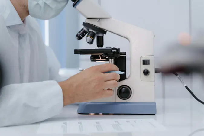
In an exciting breakthrough, researchers at MIT have developed a game-changing technique to visualize nanoscale structures within cells, making high-resolution imaging far more accessible. Traditionally, super-resolution microscopy, which is often expensive and requires specialized facilities, has been the go-to method for such imaging. However, by expanding tissue samples prior to imaging, the MIT team has enabled researchers to achieve nanoscale resolution using standard light microscopes.
20-Fold Expansion in Just One Step
The latest advancement allows researchers to expand tissue samples a staggering 20-fold in just a single step, streamlining the process. "This democratizes imaging," declares Laura Kiessling, Novartis Professor of Chemistry at MIT. "With this method, labs can now visualize previously indistinguishable structures using more affordable equipment.”
The significance of this resolution—approximately 20 nanometers—cannot be overstated. At this scale, scientists can successfully observe essential organelles such as mitochondria and intricate structures like protein clusters that were once invisible to conventional imaging methods. Edward Boyden, who holds multiple professorships at MIT, remarked, “The building blocks of life—biomolecules, genes, and their products—operate at nanoscale distances.”
The Origin of the Technique
Boyden's lab originally developed expansion microscopy in 2015 by embedding tissue into an absorbent polymer and disrupting the proteins that held the tissue together. This initial method achieved a 4-fold tissue expansion, enabling scientists to image at a resolution of 70 nanometers. Subsequent modifications introduced a two-step 20-fold expansion process, improving resolution but complicating the procedure.
The latest study, published in *Nature Methods*, has achieved the same 20-fold expansion in a single pioneering step, greatly simplifying the workflow needed for high-resolution imaging.
How It Works: A Closer Look at the Gel
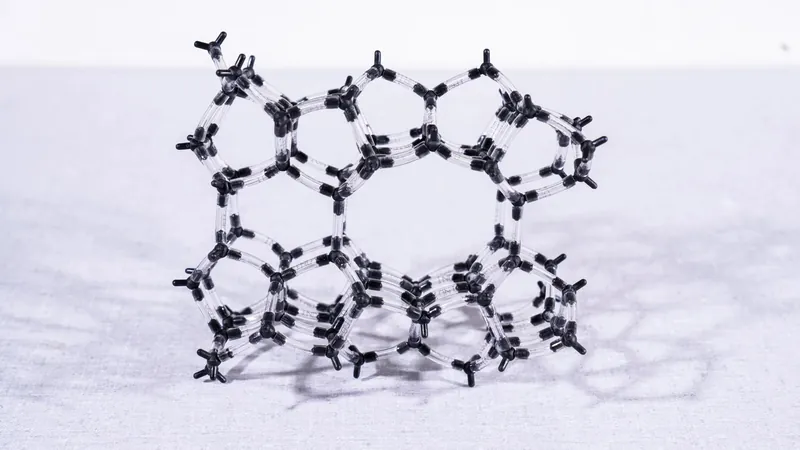
To create this new gel for expansion, the team utilized a combination of N,N-dimethylacrylamide (DMAA) and sodium acrylate. This innovative approach utilizes spontaneous crosslinking for robustness, allowing the gel to maintain its integrity while being expanded significantly. By optimizing the polymerization process and removing oxygen from the solution before gelation, researchers further enhanced the gel’s mechanical properties, enabling the ambitious 20-fold expansion.
This new method may require a bit more sample preparation compared to existing super-resolution techniques, but it greatly simplifies the subsequent imaging process, especially for 3D analyses. “We’ve outlined a detailed protocol in the study to make it easy for other researchers to replicate,” adds Tay Won Shin, one of the lead authors.
Unraveling the Mysteries of Brain and Cancer Cells
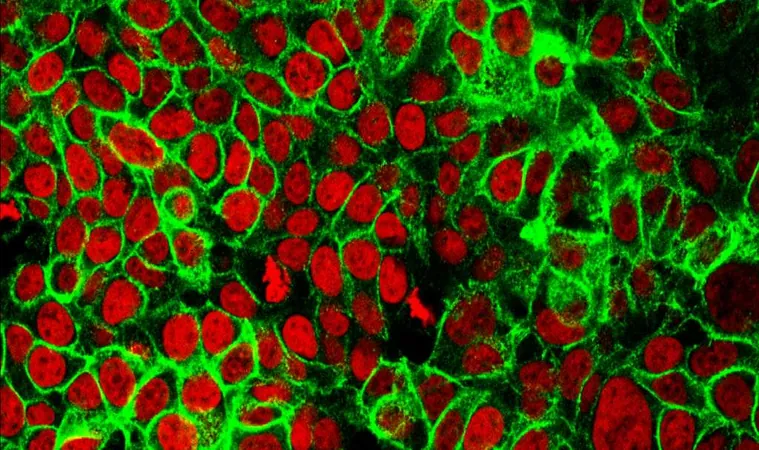
The applications of this technique are vast. In studies of brain cells, researchers successfully imaged structures called synaptic nanocolumns—vital clusters of proteins integral to neuron communication. They also explored cancer cells by observing microtubules, mitochondria, and even the organization of nuclear pore complexes.
Moreover, this method has the potential to shed light on glycan structures—carbohydrates on cell surfaces that mediate interactions with the environment. The implications are profound, as scientists could effectively visualize tumor cells, unveiling the organization of proteins within them with unprecedented ease.
The Future of Nanoscale Imaging in Biology Labs
The researchers anticipate that any biology lab should be able to employ this cost-effective technique with readily available chemicals and common equipment, such as confocal microscopes. “Our goal is for everyday biology labs to adopt this protocol using their existing instruments,” Wang explains. “With this technology, even traditional labs can achieve the resolution previously only possible with high-end, specialized microscopes.”
This groundbreaking work represents a significant leap forward in the realm of biological imaging, setting the stage for new discoveries that could unravel the complexities of life at the molecular level.

 Brasil (PT)
 Canada (EN)
 Chile (ES)
 España (ES)
 France (FR)
 Hong Kong (EN)
 Italia (IT)
 日本 (JA)
 Magyarország (HU)
 Norge (NO)
 Polska (PL)
 Schweiz (DE)
 Singapore (EN)
 Sverige (SV)
 Suomi (FI)
 Türkiye (TR)