Shocking Discovery: Nearly 44% of Interstitial Lung Abnormalities Go Unreported in CT Scans!
2024-11-20
Author: John Tan
A Study Overview
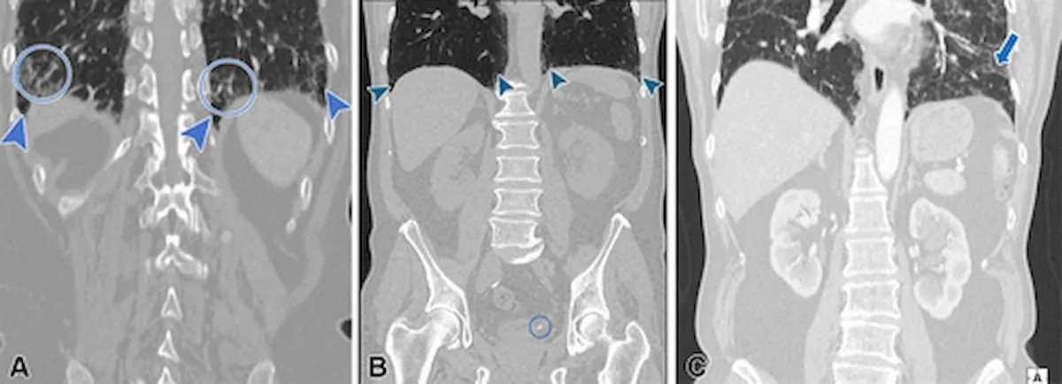
A groundbreaking study has revealed that a staggering proportion of interstitial lung abnormalities (ILAs) identified in computed tomography (CT) scans are being overlooked in patient reports. This shocking oversight could have serious implications for patient care and treatment outcomes.
Study Details
The research, recently published in the esteemed journal *Radiology*, involved an extensive retrospective analysis of 21,118 patients—most notably, with a median age of 72—who underwent abdominal or thoracoabdominal CT scans. The findings are alarming: ILAs were detected in 362 patients (approximately 1.7%), but nearly 44% of these abnormalities were not mentioned in the original CT reports examined.
Expert Insight
Dr. Nicola Sverzellati, the lead study author from the University of Parma in Italy, emphasized that the oversight can stem from a disconnect between the clinicians' focus on specific abdominal conditions and the subtle, yet critical, indicators of lung health during the reporting process. The study showed particularly concerning trends; ILAs were underreported at nearly three times the rate in abdominal CTs (72.7%) compared to thoracoabdominal CTs (29.3%).
Concerns About Unreported ILAs
The data highlighted that many of these unreported ILAs were of significant concern: over 41% were non-subpleural ILAs, while a staggering 55% of subpleural non-fibrotic ILAs also went unreported. Fibrotic ILAs—those with scarring in the lungs—were present in over 61% of patients with diagnosed ILAs and are especially critical because they were linked to a fourfold increase in mortality risk from respiratory causes.
What Does This Mean for Patients?
1. **High Underreporting Rates:** Nearly 44% of ILAs are being missed, which could delay crucial medical interventions. 2. **Clinical and Technical Shortcomings:** The lack of specific imaging techniques (like parenchymal window reconstruction) and challenges in identifying subtle lung changes, particularly in older patients, may contribute to these alarming statistics. 3. **Mortality Risks:** Fibrotic ILAs greatly increase the risk of death from respiratory diseases, underlining the urgent need for physicians to improve detection and reporting practices.
Conclusion
The implications of these findings are profound. Patients with fibrotic changes could face a significantly higher risk of mortality, emphasizing the importance of comprehensive reporting in CT scan results. This study serves as a wake-up call to the medical community, highlighting critical gaps that must be addressed to enhance patient outcomes.
As healthcare evolves, the integration of advanced imaging technologies, including artificial intelligence (AI) models, could provide solutions for improving detection rates of interstitial lung abnormalities, ultimately saving lives and enhancing care.
In conclusion, the study sheds light on a vital issue within radiology and advocates for better practices to ensure every patient's lung health is properly evaluated. Don't let your health be compromised—always discuss your CT scan results thoroughly with your healthcare provider!


 Brasil (PT)
 Canada (EN)
 Chile (ES)
 España (ES)
 France (FR)
 Hong Kong (EN)
 Italia (IT)
 日本 (JA)
 Magyarország (HU)
 Norge (NO)
 Polska (PL)
 Schweiz (DE)
 Singapore (EN)
 Sverige (SV)
 Suomi (FI)
 Türkiye (TR)