Breakthrough in Brain Organoid Selection: A Revolutionary Approach to Improve Transplant Therapy
2024-12-02
Author: Olivia
In an exciting new study, researchers led by Professor Jun Takahashi at Kyoto University have achieved a groundbreaking advance in the selection of brain organoids, a development that could transform cell replacement therapies for stroke and traumatic brain injury patients. This innovative system, outlined in the latest edition of Stem Cell Reports, combines advanced morphological analysis with single-cell gene expression techniques to ensure only the highest quality brain organoids are selected.
Organoids, which are essentially miniature, self-organizing models of organs, have emerged as an invaluable resource in medical research. They enable scientists to study organ development and simulate diseases, providing a promising avenue for generating transplantable cells and tissues. However, the inherent variability of these organoids, which often leads to the creation of non-target cell types, presents a significant challenge.
Recognizing this issue, the research team embarked on a mission to devise a non-destructive evaluation system to effectively identify the best candidates for transplantation. Their focus was on cerebral cortical organoids—structures that mimic the brain's cortex—crucial for developing therapies after neurological injuries.
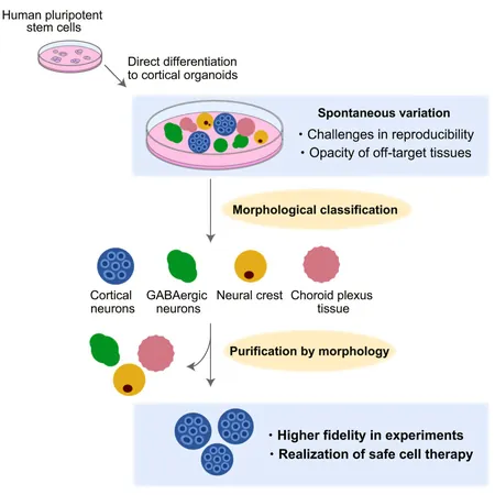
In their method, the researchers categorized cerebral cortical organoids into seven distinct types based on their morphological characteristics. Utilizing single-cell RNA sequencing (scRNA-seq), they carried out an extensive gene expression profiling that enabled them to annotate the identity of individual cells within each organoid group. This approach revealed a diverse array of neuronal subtypes, including both cortical and GABAergic neurons, alongside other cell types like fibroblasts and endothelial cells.
Notably, the scientists discovered a strong correlation between an organoid's morphology and its cellular composition. For example, organoids exhibiting structural features resembling rosettes contained a majority of cortical neurons, while those characterized by low transparency had a higher concentration of GABAergic neurons. This discovery is crucial for future therapies, as having a reliable way to predict cell types based on organoid appearance can streamline the selection process.
Further validation of these findings involved RNA microarray analysis, confirming the similarities in gene expression profiles across different organoid groups. Crucially, this reproducibility was observed in organoids produced from different cell lines, underscoring the robustness of their classification method.
With a keen interest in employing these brain organoids for cell replacement therapies, the research team specifically emphasized those with rosette-like structures. Their scRNA-seq analysis revealed that these targeted organoids consistently produced desired cortical neurons, thus significantly minimizing the risk of transplanting unintended cell types.
This revolutionary approach not only simplifies the selection process of cerebral cortical organoids but also promises a future where transplants are both safer and more effective. By using visual characteristics to guide selection, the researchers have set the stage for enhanced transplantation methodologies and improved patient outcomes in regenerative medicine.
As organoid technology continues to evolve, the implications of this work could extend far beyond brain therapies, potentially paving the way for solutions to a multitude of conditions requiring tissue regeneration. The pursuit of understanding and harnessing organoid capabilities holds vast potential for reshaping the landscape of modern medicine.


 Brasil (PT)
 Canada (EN)
 Chile (ES)
 España (ES)
 France (FR)
 Hong Kong (EN)
 Italia (IT)
 日本 (JA)
 Magyarország (HU)
 Norge (NO)
 Polska (PL)
 Schweiz (DE)
 Singapore (EN)
 Sverige (SV)
 Suomi (FI)
 Türkiye (TR)